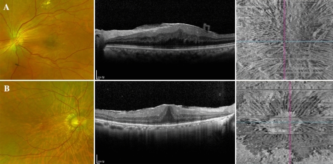
Figure 2.
Representative cases from macular pucker (A) and idiopathic ERM (B) groups. A patient of macular pucker group (A) with a history of barrier laser in the past due to a retinal break showed turbidity on fundus photography and eccentricity on the en-face OCT image. R1/R2 ratio was 6.7 and ERM extent was 12.8 mm2. A patient of idiopathic ERM with no remarkable history showed a clear ERM on fundus photography. However, an ERM was confirmed in the OCT and a concentric appearance was seen on the en-face OCT image. R1/R2 ratio was 1.9 and ERM extent was 1.8 mm2.

