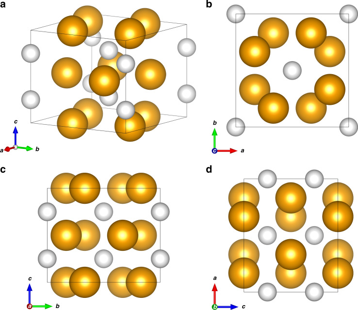
Fig. 4. Crystal structure of the (PH3)2H2 vdW compound.
Crystal structure of the (PH3)2H2 vdW compound at 5.5 GPa and room T (a) with views along the c (b), a (c), and b (d) crystal axes. The orange spheres represent the PH3 molecules, whereas the white spheres represent the H2 molecules. The size of the spheres has been drawn by estimating their radius as the sum of the P–H bond length (1.421 Å)2 and H van der Waals radius (1.20 Å)49 in the case of PH3 (2.62 Å) and as the sum of the H van der Waals radius (1.20 Å)49 and half of the H–H distance (0.742 Å)74 in the case of H2 (1.571 Å).

