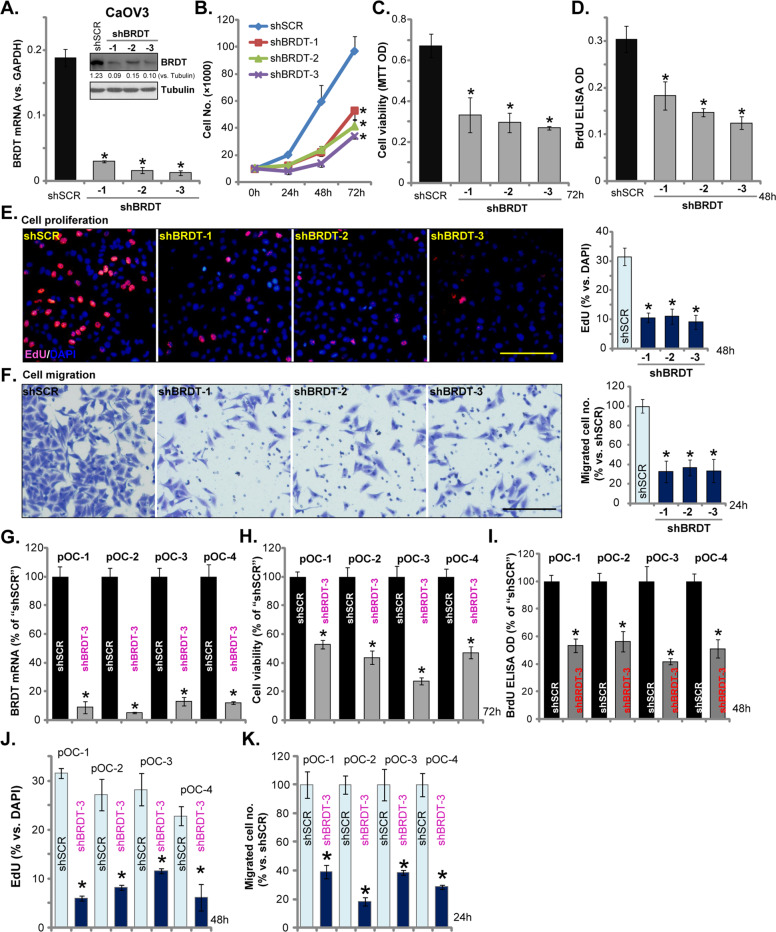
Fig. 2. BRDT shRNA inhibits ovarian cancer cell survival, growth, proliferation and migration.
CaOV3 cells (A–F) or the primary human ovarian cancer cells (“pOC-1/-2/-3/-4”) (G–K) were transfected with scramble control shRNA lentivirus (“shSCR”) or the BRDT shRNA lentivirus (“shBRDT-1/-2/-3”, with different shRNA sequence) for 24 h, stable cells were selected by puromycin. BRDT mRNA or protein expression was shown (A and G); Cell growth (B), viability (MTT assay, C and H), as well as proliferation (BrdU ELISA and EdU staining assays, D, E, I and J), and cell migration (“Transwell” assays, F and K) were tested by the appropriate assays, results were quantified. For the functional assays, the exact same amount of viable cells with different genetic modifications were initially (at 0 h) seeded into each well/dish and cultured for applied time periods (same for all Figures). BRDT protein expression was quantified and normalized to Tubulin (A). For each assay, n = 5 (five dishes or wells). *P < 0.05 vs. “shSCR” cells. Experiments in this figure were repeated five times, with similar results obtained. Scale Bar = 100 μm (E and F).

