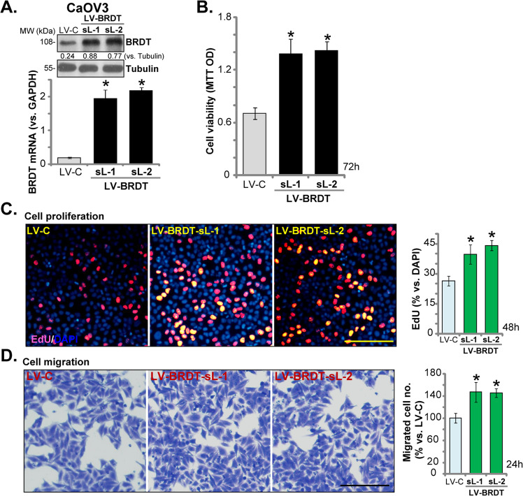
Fig. 5. BRDT overexpression promotes ovarian cancer proliferation and migration.
CaOV3 cells were transfected with lentiviral BRDT expression construct (“LV-BRDT”) or the empty vector (“LV-C”), stable cells were selected by puromycin. BRDT mRNA and protein expression (A), cell viability (MTT OD, B), proliferation (by recording EdU-positive nuclei ratio, C) and migration (“Transwell” assays, D) were tested similarly. BRDT protein expression was quantified and normalized to Tubulin (A). For each assay, n = 5 (five dishes or wells). *P < 0.05 vs. “LV-C” cells. Experiments in this figure were repeated three times, with similar results obtained. Scale Bar = 100 μm (C and D).

