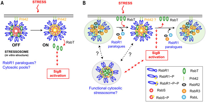
Figure 7.
Tentative model depicting the dynamics of interaction among the stressosome proteins analysed in this study. (A) Model proposed for stress response based on crystallographic data obtained in vitro with purified proteins in L. monocytogenes16; (B) Model integrating the observations obtained in vivo from L. monocytogenes about the distribution and interaction of distinct stressosome proteins. Note the putative negative role assigned to RsbR1 paralogues to prevent the formation of a stable RsbR1-RsbS-RsbT complex, which could be only transiently formed upon stress. The association of RsbT with the membrane is depicted as a direct interaction, although it could be indirect since it lacks defined hydrophobic domains. The model also highlights the presence of a large pools of cytosolic RsbR1, RsbS and RsbT, with the possibility of functional stressosomes in this location capable of activating SigB. See text for details.

