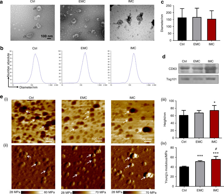
Fig. 3.
Characterization of extracellular vesicles (EVs) derived from macrophages seeded on mineralized collagen in vitro. a TEM morphology of extracellular vesicles. b Corresponding particle size and distribution analysis of the samples in a. c Semiquantification of extracellular vesicle diameter. d Western blotting of EV surface markers. e (i) AFM phase images of EVs; (ii) Corresponding modulus mapping images of EVs; (iii) Semiquantification of EV height; (iv) Semiquantification of EV modulus. Arrows: EVs; Ctrl: normal macrophage-derived sEVs. *P < 0.05 vs Ctrl, ***P < 0.001 vs Ctrl, #P < 0.05 vs EMC

