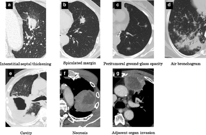
Figure 1.

Representative graphics of morphological characteristics of lung cancer assessed by computer tomography scan. (a) Interstitial septal thickening: the surrounding bronchial vascular bundle of the left upper lobe lesion presents interstitial septal thickening. (b) Spiculated margin: the fluffy shadow projects from the tumor, reaching the interlobar pleura. (c) Peritumoral ground‐glass opacity: tumors are surrounded by a region like ground‐glass. (d) Air bronchogram: in the infiltrative shadow, radiolucent shadows of the bronchus are observed. (e) Cavity: the right lower lobe lesion was a cavitary lesion. (f) Necrosis: low‐density area in the left upper lobe tumor suggests the presence of necrotic materials. (g) Adjacent organ invasion: left upper lobe tumor invades the chest wall.
