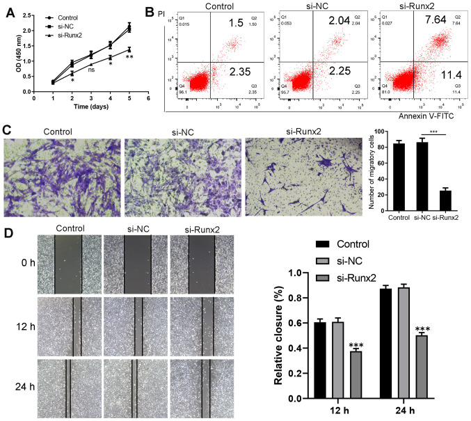
Figure 5.
Runx2 knockdown inhibits the migration and proliferation, and promotes the apoptosis of HKFs. (A) Cell Counting Kit-8 assay revealed that the proliferation of HKFs was inhibited following Runx2 knockdown. *P<0.05, **P<0.01 vs. si-NC (n=3). (B) Promotion of apoptosis following the transfection of HKFs with si-Runx2, as shown by flow cytometry using an Annexin V-FITC/PI assay. The lower left quadrant (Annexin V-/PI-) represents live cells, the lower right quadrant (Annexin V+/PI-) represents early apoptotic stage cells, the upper right quadrant (Annexin V+/PI+) represents late apoptotic stage or necrotic cells. (C) ***P<0.001 si-Runx2 vs. si-NC (n=3). (D) Evaluation of cell migration using a wound healing assay (magnification, ×100). 12 h ***P<0.001 si-Runx2 vs. si-NC (n=3); 24 h ***P<0.001 si-Runx2 vs. si-NC (n=3). Data are presented as the mean ± SD of three independent experiments. HKFs, human keloid fibroblasts; si, small interfering RNA; NC, negative control; Runx2, Runt-related transcription factor 2; PI, propidium iodide; OD, optical density.

