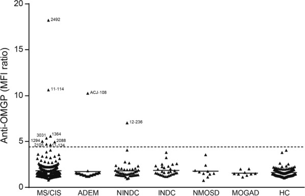
Fig. 1.

Identification of patients with Abs to OMGP. A total of 588 sera diluted 1:50 from seven cohorts were screened for OMGP autoantibodies using a cell-based assay (OMGP-TM) displaying OMGP with a transmembrane anchor. The following groups were analyzed: multiple sclerosis/clinical isolated syndrome (MS/CIS, n = 352), acute disseminated encephalomyelitis (ADEM, n = 28), non inflammatory neurological disease control (NINDC, n = 45), inflammatory neurological disease control (INDC, n = 30), neuromyelitis optica spectrum disorders (NMOSD, n = 10), MOG antibody-associated disease (MOGAD, n = 9) and healthy controls (HCs, n = 114). For the cut off evaluation, HCs were measured twice, except ten HCs samples coming from the Swedish cohort were analyzed once. The horizontal line at 4.4 represents the cut-off as mean plus 6 SDs of the HC cohort. From patients above the indicated threshold, the symbols show mean value of minimum two replicates. The numbers next to the symbols of the positive patients are the internal code numbers. Clinical details of these patients are in Table S2. ACJ-108 is a child with ADEM, patient 12–236 was diagnosed as psychosis, all other positive patients had MS. Index patient 2492 served as daily control and the value shown is the mean of 30 replicates. The raw data of the anti-OMGP reactivity of patient 2492 is shown in Fig. 2, of all other patients scored positive in Fig. S2
