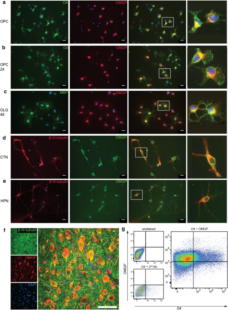
Fig. 4.
OMGP is displayed by neurons, immature and mature oligodendrocytes. Primary mouse oligodendrocyte precursor cells (OPC) (a) were differentiated for 24 h (b) or 48 h (c). A double staining was performed for OMGP (22H6) with the early oligodendroglial marker O4 (a, b), or MBP indicating differentiated oligodendrocytes (c). d Mouse cortical neurons (CTN) as well as (e) hippocampal neurons (HPN) were double-stained with the neuron-marker β-III-tubulin and OMGP (22H6). (a–e) scale bars represent 20 µm and white rectangles mark the zoomed area. (F) Spinal cord tissue sections of 55 μm were stained with anti-OMGP (22H6, red) and β-III-tubulin (green) for visualization in grey matter. Images are stacks from confocal microscopy with 60x magnification and white scale bar indicates 50 μm. g Human oligodendrocytes were double-stained with anti-OMGP (22H6-hIgG1) and O4. Quadrants were set with human IgG and secondary Abs as control

