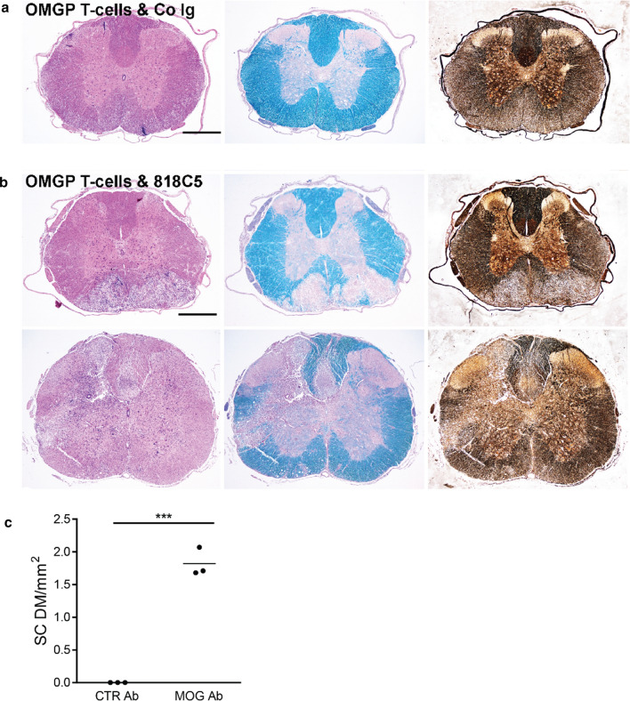
Fig. 6.
OMGP-specific T cells pave the way for anti-MOG mediated demyelination in gray and white matter of the spinal cord. Lewis rats were injected with OMGP-specific T cells, 2 days later either a control Ig (IvIg) (a) or the MOG-Ab 8-18C5-hIgG1 (b) was given and after 3 more days, the animals were sacrificed. Cross sections of the spinal cord were stained with H&E (left), LFB (middle), or Bielschowsky’s Silver Staining (right). b T cells injected together with MOG antibody (8-18C5-hIgG1), demyelination and neuronal destruction shown by Bielschowsky’s staining. Large demyelinating areas are seen in the white matter (upper part of b) and in white plus gray matter (lower part of b) along with axonal injury. Scale bar represents 1 mm. c Spinal cord (SC) demyelination (DM) was quantified from the LFB staining. The area of DM is shown in mm2 per SC section. (p < 0.001 ***; t-test)

