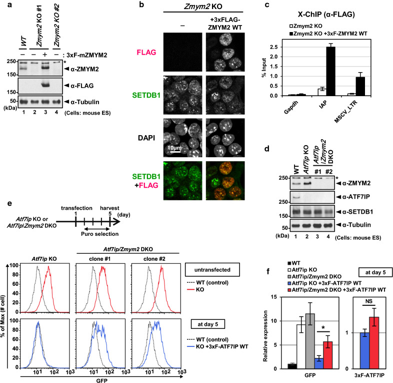
Fig. 4.
ZMYM2 partly mediates efficient re-silencing of MSCV-GFP reporter by ATF7IP. a WB analysis confirms no expression of ZMYM2 in Zmym2 KO cell lines and shows an exogenous expression of 3xFLAG-ZMYM2. b IF analysis shows the co-localization of 3xFLAG-ZMYM2 with SETDB1’s nuclear foci. c X-ChIP analysis with anti-FLAG antibody at the indicated genomic loci in Zmym2 KO mESCs and the Zmym2 KO cells rescued by 3xFLAG-ZMYM2. Gapdh gene was used as a negative control. The ChIP enrichment levels are shown as mean ± SEM; n = 3 from three experiments. d WB analysis confirms no expression of both ATF7IP and ZMYM2 in Atf7ip/Zmym2 DKO cells. e MSCV-GFP expression was analyzed by flow cytometric analysis at day 5 after the transfection. Atf7ip/Zmym2 DKO cells show higher expression of GFP, compared to the parental WT cells. f RT-qPCR analysis was performed with samples collected at day 5 after the transfection. RNA expression was normalized to Hprt expression and is shown relative to the level in WT cells (left) or Atf7ip KO cells transfected with 3xFLAG-ATF7IP WT. Data are mean ± SEM; n = 4 from four experiments. NS: P > 0.05, *P < 0.05 by unpaired Student’s t test

