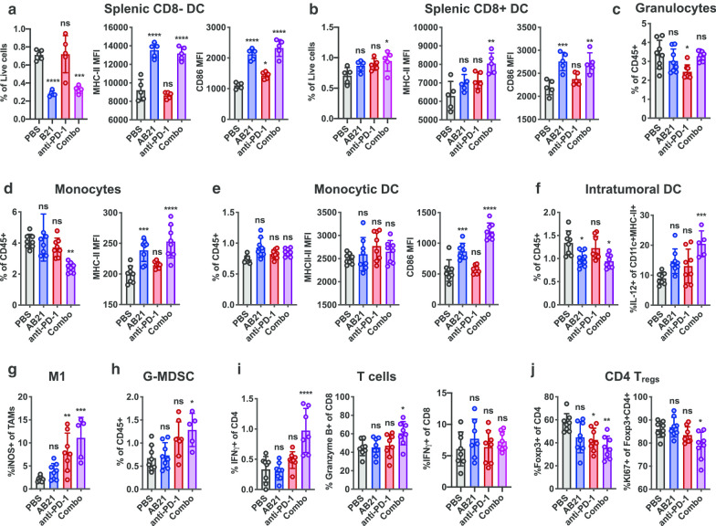
Fig. 6.
Characterization of the lymphoid and myeloid compartments of tumors and spleen. MC38 colon carcinoma cells were implanted subcutaneously in C57BL/6 mice. Mice with established tumors were randomized and treated intraperitoneally with PBS, AB21, anti-PD-1 or combo (AB21 + anti-PD-1). a, b Quantifications of splenic CD8− and CD8+ CD11chiMHCII+ DCs as a proportion of live cells and expression of costimulatory molecules in the spleen. c–e Quantification of granuloctyes (Gr1midCD11b+), monocytes (Gr1hiCD11b+) and monocytic DCs (GR1hiCD11c+CD11b+MHC-II+) as a proportion of live CD45+ cells and expression of costimulatory molecules in the spleen. f Quantification of DCs (CD11c+MHC-II+) as a proportion of live CD45+ cells and IL-12+ DCs in the tumor. g Quantification of iNOS+ expressing cells as a proportion of total TAMs (CD11b+Gr1−MHC-II+) in the tumor. h Quantification of granulocytic monocyte derived suppressor cells, G-MDSCs, (CD11b+Gr1mid) as a proportion of live CD45+ cells in the tumor. i Quantification of IFNg+ cells as a proportion of CD4 and CD8 and Granzyme B as a proportion of CD8 in the tumor. j Quantification of Foxp3+ cells as a proportion of CD4 and Ki67+ cells as of Foxp3+CD4+ Tregs in the tumor. Plotted as mean ± SD and analyzed by Ordinary one-way ANOVA, Tukey’s multiple comparisons test. *p < 0.05, **p < 0.01, ***p < 0.001, ****p < 0.0001, ns is not significant

