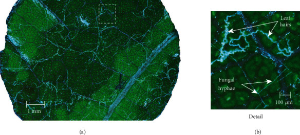
Figure 3.

Full resolution (46 megapixel) image produced through focus-stack processing of images captured in the Z-plane. (a) Leaf disk sample imaged 3 days after inoculation with Erysiphe necator conidiospores. Illumination of the live sample at near-grazing angles revealed detail of the hyaline hyphae without excessive glare from the highly reflective leaf cuticle. (b) Detailed area of image illustrating morphology of fungal hyphae and nearby leaf trichomes (hairs).
