
Figure 2.
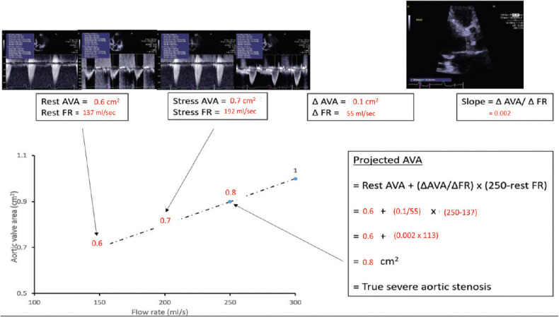
Projected aortic valve area calculation derived from resting and low-dose dobutamine echocardiography. Eighty-year-old woman with classical low-flow low-gradient severe aortic stenosis, ejection fraction of 29% and body surface area of 1.55 m2. Although stroke volume increased minimally with DSE, the flow rate increased 40% due to shortening of the ejection time, but MG and aortic valve area discordance persisted. In this example, the resting aortic valve area is 0.6 cm2 and the flow rate is 137 mL/s. The same measurements obtained during inotropic stress with low dose dobutamine give an aortic valve area of 0.7 cm2 and flow rate of 192 mL/s. Therefore, the flow rate has not normalized to at least 250 mL/s. The rate of increase in aortic valve area per unit change in flow rate is then derived from the two sets of data dividing the change in aortic valve area by the change in flow rate from rest to stress (slope of the line) = 0.002. Accordingly, the projected aortic valve area at the normalized flow rate equates to 0.8 cm2, indicating true severe aortic stenosis