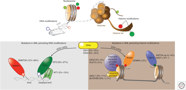
Figure 1.
Frequent mutations of epigenetic regulators in acute myeloid leukemia (AML). Upper panel illustrates epigenetic modifications on the DNA and histone layer of the epigenome. Lower panel shows gene mutations that impact DNA and/or histone modifications in at least 1% of AML cases. Ranges of reported mutation frequency are indicated in parentheses (Abbas et al. 2010; Marcucci et al. 2010, 2012; Paschka et al. 2010; Hollink et al. 2011; Shen et al. 2011; Gaidzik et al. 2012; Patel et al. 2012; Weissmann et al. 2012; Cancer Genome Atlas Research 2013; Gao et al. 2013; Metzeler et al. 2016; Papaemmanuil et al. 2016; Terada et al. 2018). The asterisk indicates frequency in adult AML. KMT, lysine methyltransferase; KDM, lysine demethylase; 2OG, 2-oxoglutarate, also called α-ketoglutarate (αKG).

