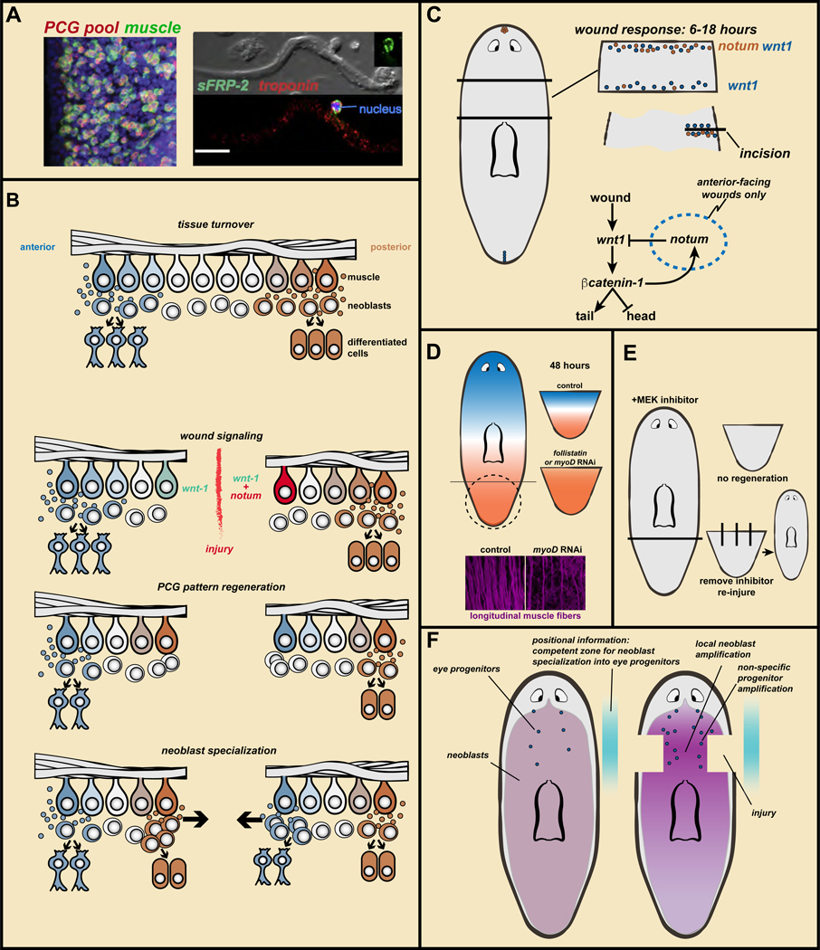
Figure 4. Wound signaling mediates the regeneration of positional information to enable regeneration of missing parts.
A. PCGs are expressed in muscle. Data from (Witchley et al., 2013). Left, a PCG RNA probe pool (red) labels almost all body-wall muscle cells (green, collagen probe). Right, sFRP-2 transcripts around a muscle cell nucleus. Bar, 20 microns. B. Regeneration model: muscle ßPCG expression, specialized neoblasts, and wound-induced re-establishment of PCG expression domains. See text for details. C. notum is preferentially wound-induced at anterior-facing wounds and wnt1 is wound-induced at all wounds. notum inhibits Wnt signaling to promote head regeneration. How notum is selectivity activated at anterior-facing wounds is unknown. D. Without PCG pattern regeneration in follistatin or myoD RNAi animals, regeneration fails to occur. This requires longitudinal muscle fibers (data from (Scimone et al., 2017)). E. MEK inhibitor treatment blocks regeneration. Removal of the inhibitor does not lead to regeneration unless new injuries are inflicted. F. Coincidence of increased neoblast proliferation with positional information leads to amplification of regional progenitor classes, even if the target tissue of those progenitors remains uninjured.

