. 2020 Oct 13;297(3):545–553. doi: 10.1148/radiol.2020200240

Figure 2:

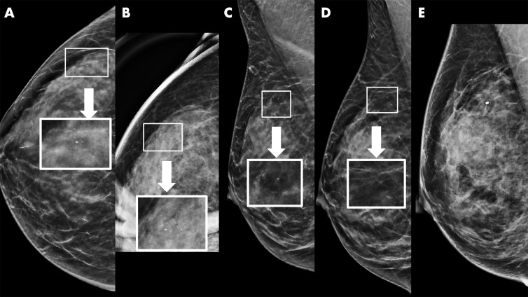
Example of synthetic imaging calcification enhancement in a 43-year-old woman who presented for screening. A, Screening mammogram obtained with synthetic imaging (craniocaudal view). New faint amorphous calcifications are seen in the outer breast. Finding was classified as Breast Imaging Reporting and Data System (BI-RADS) category 0. Inset image was obtained at ×3 magnification. B, Diagnostic mammogram, two-dimensional magnification view (craniocaudal view), confirms the presence of calcifications; however, they are of equal or less conspicuity compared with that seen with synthetic imaging. Inset image was obtained at ×3 magnification. Finding was classified as BI-RADS category 4. C, Screening mammogram obtained with synthetic imaging (mediolateral oblique view). New faint amorphous calcifications are seen in the superior breast. They are less conspicuous compared with those on the craniocaudal view in A. Finding was classified as BI-RADS category 0. Inset image was obtained at ×3 magnification. D, Diagnostic digital mammogram obtained at recall (mediolateral view). Calcifications are much less conspicuous on two-dimensional digital mediolateral view. Inset image was obtained at ×3 magnification. Finding was classified as BI-RADS category 4. E, Diagnostic mammogram obtained with two-dimensional imaging after biopsy (digital mediolateral view). The clip is in appropriate location. Pathologic examination showed ductal carcinoma in situ, intermediate grade.

Example of synthetic imaging calcification enhancement in a 43-year-old woman who presented for screening. A, Screening mammogram obtained with synthetic imaging (craniocaudal view). New faint amorphous calcifications are seen in the outer breast. Finding was classified as Breast Imaging Reporting and Data System (BI-RADS) category 0. Inset image was obtained at ×3 magnification. B, Diagnostic mammogram, two-dimensional magnification view (craniocaudal view), confirms the presence of calcifications; however, they are of equal or less conspicuity compared with that seen with synthetic imaging. Inset image was obtained at ×3 magnification. Finding was classified as BI-RADS category 4. C, Screening mammogram obtained with synthetic imaging (mediolateral oblique view). New faint amorphous calcifications are seen in the superior breast. They are less conspicuous compared with those on the craniocaudal view in A. Finding was classified as BI-RADS category 0. Inset image was obtained at ×3 magnification. D, Diagnostic digital mammogram obtained at recall (mediolateral view). Calcifications are much less conspicuous on two-dimensional digital mediolateral view. Inset image was obtained at ×3 magnification. Finding was classified as BI-RADS category 4. E, Diagnostic mammogram obtained with two-dimensional imaging after biopsy (digital mediolateral view). The clip is in appropriate location. Pathologic examination showed ductal carcinoma in situ, intermediate grade.