. 2020 Oct 20;297(3):503–512. doi: 10.1148/radiol.2020200771

Figure 2c:

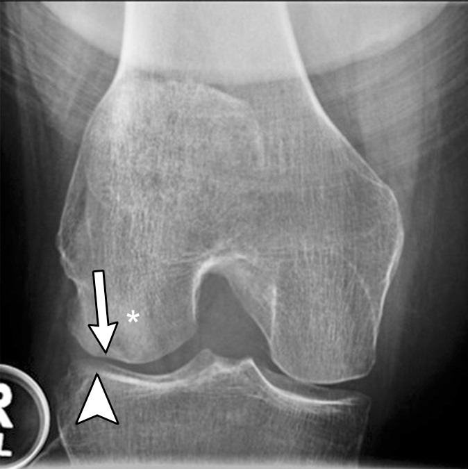
Subchondral insufficiency fracture in a 61-year-old woman with severe knee pain unrelated to trauma referred to radiology for intra-articular corticosteroid (IACS) injection. (a) Anteroposterior radiograph of the right knee obtained the day of the IACS injection shows mild osteoarthritis (OA) with small osteophytes of the lateral tibia and femur (arrows) and no joint space narrowing. (b) Coronal fat-suppressed proton density-weighted MRI performed 1 month after the IACS injection shows subchondral insufficiency fracture (arrow) with extensive bone marrow edema of the lateral femoral condyle (*) and adjacent soft tissue edema. There is also a severe lateral meniscus extrusion (arrowhead). (c) Repeat radiograph of the right knee 3 months later shows the subchondral insufficiency fracture with collapse of the articular contour of the lateral femoral condyle (arrow) surrounded by bone sclerosis (*) and lateral tibiofemoral joint space narrowing (arrowhead) likely secondary to the severe lateral meniscal subluxation. A normal or mild OA baseline radiograph in a patient with severe joint pain as in this case should trigger a preprocedural MRI to depict occult findings of clinical relevance such as subchondral insufficiency fracture.

Subchondral insufficiency fracture in a 61-year-old woman with severe knee pain unrelated to trauma referred to radiology for intra-articular corticosteroid (IACS) injection. (a) Anteroposterior radiograph of the right knee obtained the day of the IACS injection shows mild osteoarthritis (OA) with small osteophytes of the lateral tibia and femur (arrows) and no joint space narrowing. (b) Coronal fat-suppressed proton density-weighted MRI performed 1 month after the IACS injection shows subchondral insufficiency fracture (arrow) with extensive bone marrow edema of the lateral femoral condyle (*) and adjacent soft tissue edema. There is also a severe lateral meniscus extrusion (arrowhead). (c) Repeat radiograph of the right knee 3 months later shows the subchondral insufficiency fracture with collapse of the articular contour of the lateral femoral condyle (arrow) surrounded by bone sclerosis (*) and lateral tibiofemoral joint space narrowing (arrowhead) likely secondary to the severe lateral meniscal subluxation. A normal or mild OA baseline radiograph in a patient with severe joint pain as in this case should trigger a preprocedural MRI to depict occult findings of clinical relevance such as subchondral insufficiency fracture.