. 2020 Oct 20;297(3):503–512. doi: 10.1148/radiol.2020200771

Figure 3b:

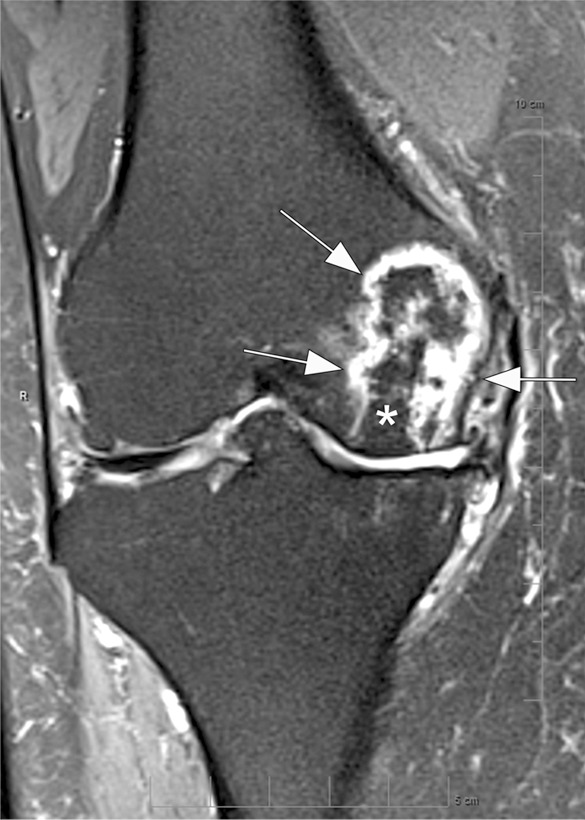
Osteonecrosis in a 59-year-old man referred to radiology for intra-articular corticosteroid (IACS) injection. (a) Anteroposterior radiograph of the right knee shows severe osteoarthritis with bone on bone appearance (arrows) and large definite osteophytes (arrowheads). There is a subchondral sclerosis of the medial femoral condyle (*), which is expected in advanced osteoarthritis. No sign of subchondral insufficiency fracture or osteonecrosis. Patient was experiencing an acute episode of pain exacerbation at time of presentation. (b) Coronal fat-suppressed intermediate-weighted MRI was performed before the IACS injection and discloses a large area of osteonecrosis of the medial femoral condyle (arrows) with pathognomonic serpiginous demarcation and fat-equivalent center of lesion (*). No collapse of the articular contour is seen. There is attrition (ie, surface remodeling) as part of the advanced osteoarthritis process. IACS injection was not performed.

Osteonecrosis in a 59-year-old man referred to radiology for intra-articular corticosteroid (IACS) injection. (a) Anteroposterior radiograph of the right knee shows severe osteoarthritis with bone on bone appearance (arrows) and large definite osteophytes (arrowheads). There is a subchondral sclerosis of the medial femoral condyle (*), which is expected in advanced osteoarthritis. No sign of subchondral insufficiency fracture or osteonecrosis. Patient was experiencing an acute episode of pain exacerbation at time of presentation. (b) Coronal fat-suppressed intermediate-weighted MRI was performed before the IACS injection and discloses a large area of osteonecrosis of the medial femoral condyle (arrows) with pathognomonic serpiginous demarcation and fat-equivalent center of lesion (*). No collapse of the articular contour is seen. There is attrition (ie, surface remodeling) as part of the advanced osteoarthritis process. IACS injection was not performed.