. 2020 Oct 20;297(3):503–512. doi: 10.1148/radiol.2020200771

Figure 4d:

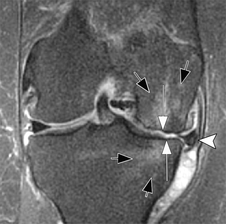
Rapid progressive osteoarthritis (RPOA) type 1 in a 52-year-old man referred to radiology for intra-articular corticosteroid (IACS) injection. (a) Anteroposterior radiograph of the right knee shows mild osteoarthritis with definite osteophytes (arrows) and minimal joint space narrowing of the medial tibiofemoral joint (arrowheads). (b) Baseline coronal fat-suppressed intermediate-weighted MRI scan confirms the osteophytes (black arrows) and shows diffuse cartilage loss at the medial femoral condyle (white arrow) with moderate subluxation of the medial meniscus (arrowhead). (c) Six months after the IACS injection, a repeat anteroposterior radiograph of the right knee shows severe medial tibiofemoral joint space narrowing (arrows) with loss of more than 2 mm of joint space width consistent with rapid progressive osteoarthritis type 1. (d) Coronal fat-suppressed intermediate-weighted MRI scan confirms extensive loss of cartilage at the medial femur and tibia (white arrows) and worsening of the medial meniscal subluxation (arrowhead). There is also subchondral bone marrow edema at the medial tibia and femur (black arrows).

Rapid progressive osteoarthritis (RPOA) type 1 in a 52-year-old man referred to radiology for intra-articular corticosteroid (IACS) injection. (a) Anteroposterior radiograph of the right knee shows mild osteoarthritis with definite osteophytes (arrows) and minimal joint space narrowing of the medial tibiofemoral joint (arrowheads). (b) Baseline coronal fat-suppressed intermediate-weighted MRI scan confirms the osteophytes (black arrows) and shows diffuse cartilage loss at the medial femoral condyle (white arrow) with moderate subluxation of the medial meniscus (arrowhead). (c) Six months after the IACS injection, a repeat anteroposterior radiograph of the right knee shows severe medial tibiofemoral joint space narrowing (arrows) with loss of more than 2 mm of joint space width consistent with rapid progressive osteoarthritis type 1. (d) Coronal fat-suppressed intermediate-weighted MRI scan confirms extensive loss of cartilage at the medial femur and tibia (white arrows) and worsening of the medial meniscal subluxation (arrowhead). There is also subchondral bone marrow edema at the medial tibia and femur (black arrows).