. 2020 Oct 20;297(3):503–512. doi: 10.1148/radiol.2020200771

Figure 5c:

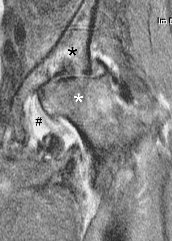
Rapid progressive osteoarthritis type 2 in a 38-year-old woman referred to radiology for intra-articular corticosteroid (IACS) injection. (a) Baseline anteroposterior radiograph of the left hip shows mild osteoarthritis with small definite osteophytes at the lateral acetabulum and femoral head (arrows) and no definite joint space narrowing. (b) Six months after the IACS injection a repeat anteroposterior radiograph of the left hip shows a complete collapse of the head of the femur with marked bone loss of the femoral head (arrow) and surface remodeling and flattening of the acetabulum (arrowheads). (c) Coronal fat-suppressed proton density-weighted MRI scan obtained on the same day demonstrates diffuse bone marrow edema of the femur and acetabulum (black and white *) and a large hip joint effusion (#) reflecting an ongoing process of marked synovial activation.

Rapid progressive osteoarthritis type 2 in a 38-year-old woman referred to radiology for intra-articular corticosteroid (IACS) injection. (a) Baseline anteroposterior radiograph of the left hip shows mild osteoarthritis with small definite osteophytes at the lateral acetabulum and femoral head (arrows) and no definite joint space narrowing. (b) Six months after the IACS injection a repeat anteroposterior radiograph of the left hip shows a complete collapse of the head of the femur with marked bone loss of the femoral head (arrow) and surface remodeling and flattening of the acetabulum (arrowheads). (c) Coronal fat-suppressed proton density-weighted MRI scan obtained on the same day demonstrates diffuse bone marrow edema of the femur and acetabulum (black and white *) and a large hip joint effusion (#) reflecting an ongoing process of marked synovial activation.