. 2020 Oct 20;297(3):503–512. doi: 10.1148/radiol.2020200771

Figure 6a:

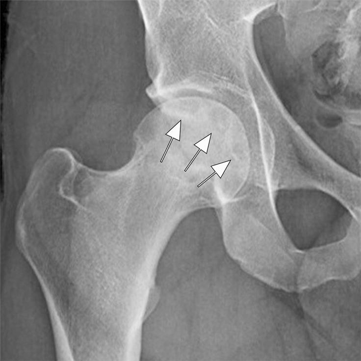
Osteonecrosis in a 29-year-old man who presented with right hip pain. (a) Anteroposterior radiograph and (b) coronal fat-suppressed proton density-weighted MRI of the right hip obtained on the same day show osteonecrosis in the right femoral head, with preserved femoral head contours (arrows). He subsequently went to the sports medicine clinic and was administered a right hip joint intra-articular corticosteroid (IACS) injection for pain. Three months later, he was referred to our institution for repeat IACS injection due to worsening pain. (c) Repeat anteroposterior right hip radiograph shows collapse of the superior femoral head articular surface (arrows). (d) Coronal reformatted CT image of the right hip confirms the collapse of the superior femoral head articular surface (arrows) and shows new hip joint space narrowing. Patient subsequently underwent right hip joint replacement.

Osteonecrosis in a 29-year-old man who presented with right hip pain. (a) Anteroposterior radiograph and (b) coronal fat-suppressed proton density-weighted MRI of the right hip obtained on the same day show osteonecrosis in the right femoral head, with preserved femoral head contours (arrows). He subsequently went to the sports medicine clinic and was administered a right hip joint intra-articular corticosteroid (IACS) injection for pain. Three months later, he was referred to our institution for repeat IACS injection due to worsening pain. (c) Repeat anteroposterior right hip radiograph shows collapse of the superior femoral head articular surface (arrows). (d) Coronal reformatted CT image of the right hip confirms the collapse of the superior femoral head articular surface (arrows) and shows new hip joint space narrowing. Patient subsequently underwent right hip joint replacement.