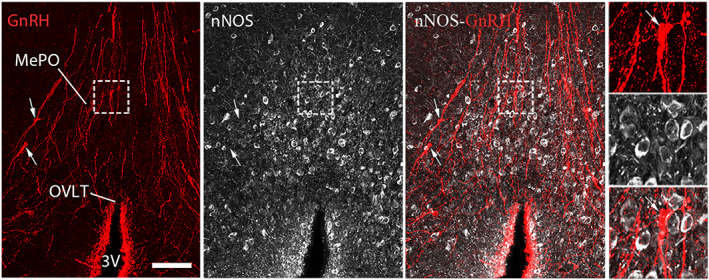
FIGURE 4.

Representative images showing nNOS and GnRH immunoreactive neurons in the preoptic region in mice. GnRH neuronal cell bodies (arrows, red) and processes (red) morphologically interact with nNOS neurons (white) in the median preoptic nucleus (MePO). However, GnRH immunoreactivity does not colocalize with nNOS immunoreactivity. OVLT, organum vasculosum of the lamina terminalis; 3V, third ventricle. Scale bar = 100 μm (25 μm in insets). Adapted from Chachlaki, Malone, et al. (2017) with permission
