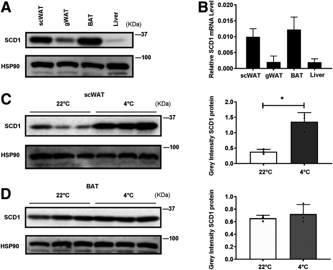
Fig. 2.
SCD1 is enriched in scWAT and BAT. It is highly induced by cold exposure in adipocytes. A: Tissue distribution of SCD1 protein expression in adipocytes of scWAT, gWAT, BAT, and liver from mice housed at RT (22°C). B: Tissue distribution of Scd1 mRNA expression (relative to 18S rRNA) in adipocytes of scWAT, gWAT, BAT, and liver from mice housed at RT. C: Western blot detection of SCD1 expression in scWAT adipocytes of mice at RT or during cold exposure (4°C) for 3 days (left panel), and the gray density of those bands (right panel) (n = 3 for each group). D: Western blot detection of SCD1 expression in BAT adipocytes of mice at RT or during cold exposure for 3 days (left panel), and the gray density of those bands (right panel) (n = 3 for each group). Statistical analysis: unpaired Student’s t-test in C and D. Data were expressed as mean ± SD. *P < 0.05, **P < 0.01, ***P < 0.001.

