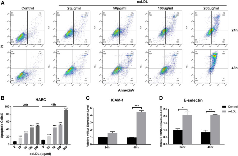
Fig. 2.
oxLDL induces inflammation and apoptosis in HAECs. A: Flow cytometry analysis of apoptotic HAECs without or with oxLDL treatment (25, 50, 100, and 200 μg/ml, respectively) after 24 and 48 h of treatment. B: Quantification of apoptotic cells after treatment with different oxLDL concentrations. C: Gene expression levels of ICAM-1 in HAECs with or without oxLDL treatment at 50 μg/ml oxLDL for 24 and 48 h. D: Gene expression levels of E-selectin in HAECs with or without oxLDL treatment at 50 μg/ml oxLDL for 24 and 48 h. Controls were untreated cells. Error bars represent standard deviations. *P < 0.05, **P < 0.01, ***P < 0.001, compared with untreated cells, one-way ANOVA followed by post hoc Dunnett’s test (B). Group pairs were compared by the Student’s t-test (C, D). Q8

