Figure 2.

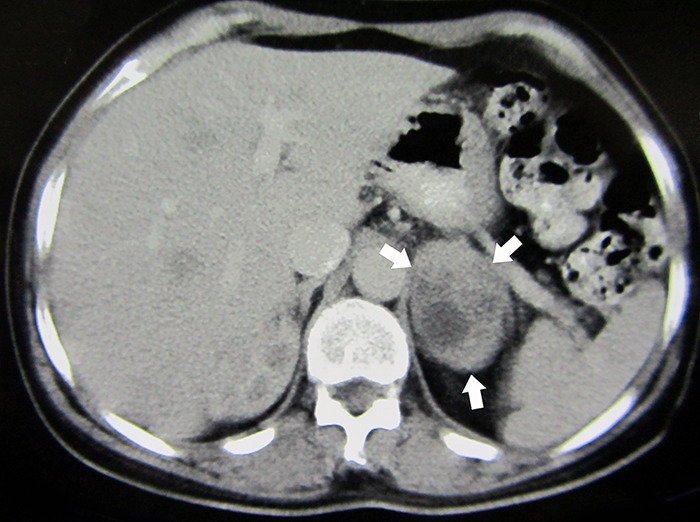
Abdominopelvic computed tomography (CT) shows a large heterogeneous solid cystic mass (60×40 mm) in the left adrenal gland (white arrows).

Abdominopelvic computed tomography (CT) shows a large heterogeneous solid cystic mass (60×40 mm) in the left adrenal gland (white arrows).