. 2020 Oct 20;9:e62043. doi: 10.7554/eLife.62043

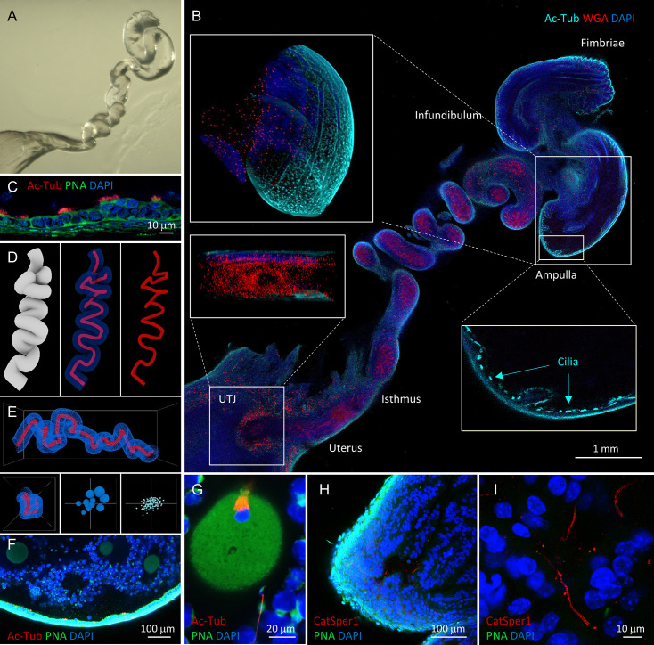
Figure 3. Tissue clearing preserves morphology of female reproductive tract and enables molecular imaging and post-processing of gametes in situ.

(A) Refractive index-matched cleared mouse female reproductive tract by CLARITY-based tissue clearing. (B) Optical imaging of the cleared female reproductive tract stained by WGA (red), Ac-Tub antibody (cyan) and DAPI (blue), 100×. Insets show cilia stained by Ac-Tub antibody in 2D (bottom right), a 3D (top left) projection of the ampulla, and a UTJ cross-section (bottom left). (C) Details of the ampullar epithelium stained by PNA (green), Ac-Tub antibody (red) and DAPI (blue), 400×. (D) 3D digital image reconstruction of the oviduct representing different 3D images rendered for oviductal surface (left) and central lumen of oviduct with (middle) or without (right) oviductal volume information. (E) Morphometric and fluorescent signal quantification analysis of the oviduct showing the morphometric meshwork representation of the 3D volumetric data from the oviduct imaging (top), the corresponding side view (bottom left) and the non-numerical visual representations of the basic volumetric (bottom middle) and fluorescent (bottom right) properties. (F) A fluorescent image showing a closer look of the cleared ampulla with oocytes (oocyte magnified in the panel G on the right-most side), 100×. (G) An oocyte with the meiotic spindle; a sperm cell is approaching the zona pellucida directly inside the ampulla, PNA (green), anti-AcTub antibody (red) and DAPI (blue), 630×. (H) A tile-scanned confocal image of the epithelium of the cleared ampulla (8 hr post-coitus) stained by anti-CatSper1 antibody (red), PNA (green), and DAPI (blue), 100×. (I) Details of the sperm stained directly inside the ampulla by anti-CatSper1 antibody (red). Two linear CatSper domains are clearly recognizable by confocal imaging. Cell nuclei are stained with DAPI (blue); acrosomes are stained with PNA (green). Images shown here are representative of at least three independent experiments. See also Figure 3—videos 37.

Figure 3.

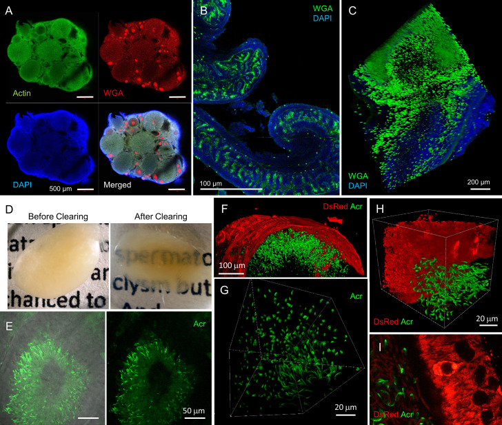
Figure 3—figure supplement 1. Multicolor 3D fluorescence in situ imaging of cleared mouse reproductive organs.

Figure 3—figure supplement 1.

(A–C) Confocal images of cleared female reproductive organs. X-CLARITY processed ovary (A); actin stained by phalloidin (green), WGA (red), and DAPI (blue). Developing oocytes show intense WGA signal. Oviduct (B) and UTJ (C, 3D rendered) from PACT-PRESTO processed female reproductive tract; stained with WGA (green) and DAPI (blue). Mucus in the lumen of the female tract is preserved (WGA, green). (D–I) Images of X-CLARITY-cleared male reproductive organs. Whole testis before (left) and after (right) clearing (D), a seminiferous tubule from the cleared testis (E), 3D surface presentations of a seminiferous tubule (F) and sperm inside the tubule (G) of SWITCH-cleared testis from Su9-DsRed2/Acr-EGFP transgenic male. (H–I) Fluorescence images of an X-CLARITY cleared epididymis from a Su9-DsRed2/Acr-EGFP male. A 3D rendered image (H) and a confocal image (I). EGFP, green; Red, DsRed. Images shown here are representative of three independent experiments. See also Figure 3—videos 17.
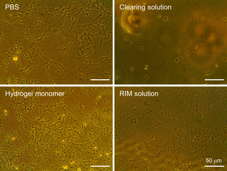
Figure 3—figure supplement 2. The effect of various components used in tissue clearing procedure on sperm cells.

Figure 3—figure supplement 2.

Uterine sperm from whole-body fixed females (3 hr post-coitus) by cardiac perfusion of hydrogel monomer were adhered to glass slides and exposed for 5 days to PBS, clearing solution, hydrogel monomer, and RIM solution. Without post-fixation and polymerization, sperm cells released from the fixed female tract were lysed and detached by clearing solution containing 4% SDS, due to incomplete fixation in the lumen and no supportive matrix to fix the cell location.
Figure 3—video 1. 3D volume imaging of a whole ovary; related to Figure 3.
Download video file (2.9MB, mp4)
(A) A movie of stitched and Z-stacked confocal slices of X-CLARITY cleared and stained ovary. (B) A 3D rendered movie from (A). Blue, DAPI; Green, Actin; Red, WGA.
Figure 3—video 2. 3D Volume imaging of the testis and epididymis; related to Figure 3.
Download video file (10.3MB, mp4)
(A) A 3D rendered movie of stitched and Z-stacked confocal slices of the testis. (B) A movie of Z-stacked confocal images of the epididymis. X-CLARITY cleared a Su9-DsRed2/Acr-EGFP male. DAPI, blue; DsRed, red; EGFP, green.
Figure 3—video 3. 3D rotational movie of whole female reproductive tract.
Download video file (1.1MB, mp4)
3D reconstruction of the cleared female reproductive tract corresponding to the Figure 3 showing voluminous uterus continuing into the oviduct and ampulla. DAPI, blue; WGA, red; Ac-Tub, cyan.
Figure 3—video 4. z-stack movie of the ampulla.
Download video file (4.2MB, mp4)
z-stack flight through video showing the ampulla with oocytes. DAPI, blue; WGA, green.
Figure 3—video 5. z-stack movie of the oviduct.
Download video file (3.4MB, mp4)
(A) 3D reconstruction of the oviductal isthmus. (B) z-stack flight-through projection. DAPI, blue; WGA, green.
Figure 3—video 6. 3D rotational movie of the UTJ.
Download video file (4.6MB, mp4)
Reconstruction of the UTJ showing 3D projections. DAPI, blue; WGA, green.
Figure 3—video 7. Moments after fertilization.
Download video file (4.5MB, mp4)
3D reconstruction of the oocyte and sperm in the cleared ampulla. (A) A flight-through z-stack video showing the oocyte with meiotic spindle and sperm stained by AcTub signal approaching the zona-pellucida. (B) The corresponding 3D projection video to (A). DAPI, blue; PNA, green; AcTub, red.