. 2020 Dec 1;9:e60226. doi: 10.7554/eLife.60226

Figure 1. A lack of supplemental glutamine in growth medium protects intracellular T. cruzi amastigotes from the cytocidal effects of ketoconazole.

(A) Dose response curves at 66 hpi of benznidazole and (B) ketoconazole treatment, in the indicated media compositions, normalized to the largest mean in each treatment group. Mean (symbols) and standard deviation shown (n = 4). (C) Microscopic counts at 66 hpi and (D) 90 hpi of the number of amastigotes per infected host cell (n = 40), medians indicated. Cartoons at top of graph indicate conditions where extracellular trypomastigotes are visible in the culture supernatant. (E) Growth media was replaced and extracellular trypomastigotes were counted every 24 hr beginning at 90 hpi (n = 2). (F) Detection of clonal outgrowth 14 days after the indicated treatments, normalized to DMSO (vehicle) treatment. Mean and standard deviation shown, circles indicates values of two independent experiments with 28 wells used per treatment within an experiment. (G) Dose response curves of glutamine in the presence of DMSO or (H) ketoconazole (5 nM). Mean and standard deviation shown (n = 3). Grey shading indicates in panel I shows the physiological range found in human plasma (800–300 uM) (Cruzat et al., 2018). Statistical comparisons between medians (C,D) were performed using a Kruskal-Wallis test with Dunn’s multiple comparisons test (****p<0.0001, ns = not significant). Comparisons of means from outgrowth (F) was performed using a two-way ANOVA with Dunnett’s multiple comparisons test (*p<0.05, **p<0.01).

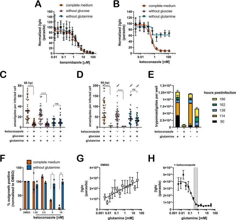
Figure 1.

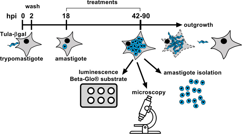
Figure 1—figure supplement 1. Experimental schematic for in vitro infection and readouts.

Figure 1—figure supplement 1.

Trypomastigotes (Tula-βgal) are incubated with mammalian host cells for 2 hr to allow invasion. Remaining extracellular parasites are subsequently removed by thorough rinsing of monolayers. Internalized parasites undergo differentiation into mature amastigotes and any treatments or media adjustments are initiated at 18 hpi prior to the first amastigote division. At indicated time points post-infection (e.g. 42–90 hpi), infected cultures have one of several fates depending on the experiment, as illustrated and described in detail in the Materials and methods.
Figure 1—figure supplement 2. Sensitivity to additional azole drugs is modulated by glutamine.

Figure 1—figure supplement 2.

(A) Dose response curves of itraconazole, (B) ravuconazole, and (C) posaconazole treatment measured at 66 hpi. Treatment including media compositions are indicated and growth is normalized to the largest mean in each data set. Mean (symbols) and standard deviation show (n = 4).
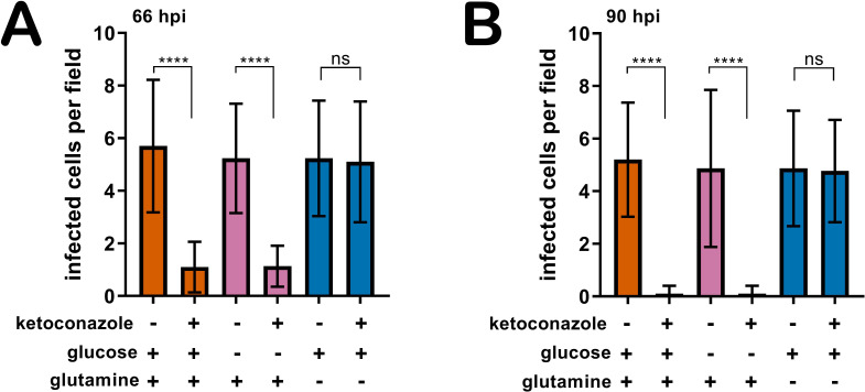
Figure 1—figure supplement 3. Removal of supplemental glutamine but not glucose maintains the proportion of infected host cells in the presence of azoles.

Figure 1—figure supplement 3.

The number of infected cells per field (n = 20) at (A) 66 hpi and (B) 90 hpi, mean and standard deviations shown. Comparisons of means (D,F) were performed using a one-way ANOVA and Bonferroni’s multiple comparisons test (****p<0.0001, ns = not significant).
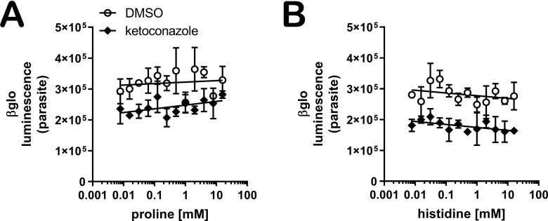
Figure 1—figure supplement 4. Proline or histidine supplementation do not sensitize amastigotes to ketoconazole in the absence of glutamine.

Figure 1—figure supplement 4.

Dose response curves of (A) proline and (B) histidine in the absence of supplemental glutamine (n = 3).
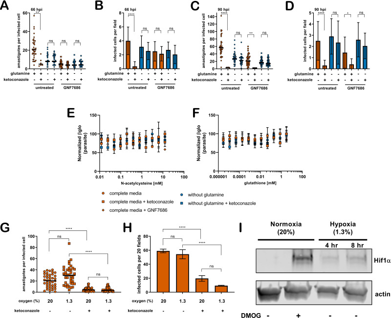
Figure 1—figure supplement 5. Slowed amastigote growth, antioxidants, or hypoxia does not prevent the cidal effects of ketoconazole.

Figure 1—figure supplement 5.

(A) Microscopic counts of amastigotes per host cell (n = 40) and (B) proportion of infected cells (n = 20) at 66 hpi following treatment at 18 hpi with ketoconazole (5 nM) and/or GNF7686 (150 nM) under the indicated conditions. (C) Microscopic counts of amastigotes per host cell (n = 40) and (D) proportion of infected cells (n = 20) at 90 hpi following treatment at 18 hpi with ketoconazole (5 nM) and/or GNF7686 (150 nM) under the indicated conditions. Statistical comparisons between medians (A,C) were performed using a Kruskal-Wallis test with Dunn’s multiple comparisons test (****p<0.0001, **p<0.01, ns = not significant). Comparisons of means (B,D) were performed using a one-way ANOVA and Bonferroni’s multiple comparisons test (****p<0.0001, *p<0.05, ns = not significant). (E) Normalized dose response of N-acetylcysteine and (F) glutathione measured at 66 hpi in the indicated treatment conditions. Mean and standard deviations are show (n = 2). (G) Microscopic counts of the number of amastigotes per infected host cell, mean indicated, (n = 40) and (H) the number of infected cells per 20 fields, mean and standard deviation shown (n = 3). Growth in complete medium under normoxia (20% atmospheric oxygen) or hypoxia (1.3% oxygen) and ketoconazole (5 nM) where indicated. (I) Western blot of uninfected whole host cell lysate. Hif1α is induced under hypoxia and in the presence of DMOG (0.8 mM for 6 hr) as a positive control.