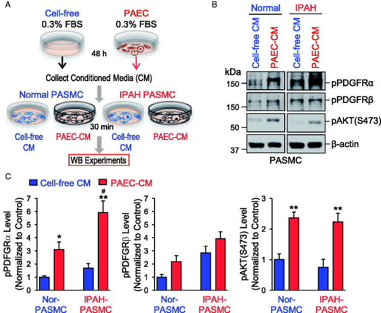
Fig. 5.
CM collected from PAEC activates PDGFRα in PASMC. (A) Schematic diagram showing how the CM was collected for Western blot experiments from cell-free controls and normal PAEC and applied to normal and IPAH PASMC to test the effects of CM on PDGFR. (B) Representative Western blot images on phosphorylated (p) PDGFRα (pPDGFRα), PDGFRβ (pPDGFRβ), and AKT (pAKT-S473) in normal and IPAH PASMC incubated for 30 min with the CM collected from cell-free controls (cell-free CM) or normal PAEC (PAEC-CM) after 48 h. (C) Summarized data (means ± SE) showing the protein expression of pPDGFRα (left panel), pPDGFRβ (middle panel), and pAKT-S473 (right panel) in normal and IPAH-PASMC incubated with cell-free CM or PAEC-CM for 30 min. *p < 0.05, **p < 0.01 versus cell-free CM; #P < 0.05 versus normal PASMC. 0.3% FBS basal medium was used for the collection. FBS: fetal bovine serum; IPAH: idiopathic PAH; PAEC: pulmonary arterial endothelial cell; pAKT: phosphorylated AKT; PASMC: pulmonary arterial smooth muscle cell; PDGFRα: platelet-derived growth factor receptor α; PDGFRβ: platelet-derived growth factor receptor β.

