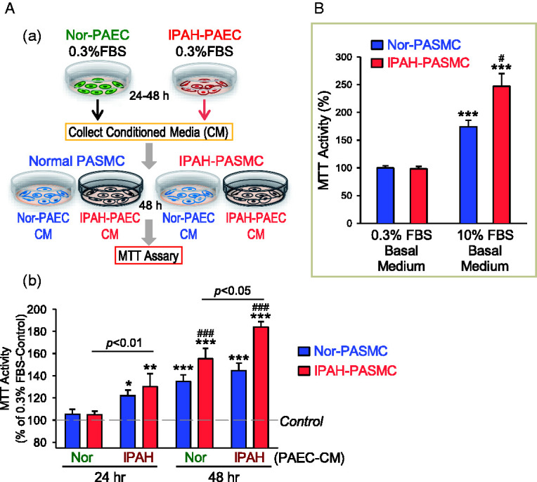
Fig. 6.

CM collected from normal and IPAH PAEC stimulates PASMC proliferation. (A) Schematic diagram (a) showing how the CM was collected for MTT experiments from normal (Nor-PAEC) and IPAH PAEC (IPAH-PAEC) and applied to PASMC isolated from normal subjects (normal PASMC) and IPAH patients (IPAH-PASMC) to test the effects of PAEC-CM on PASMC proliferation. Summarized data (b, means ± SE) showing MTT activity in normal PASMC (Nor-PASMC) and IPAH-PASMC cultured for 48 h in Nor-PAEC-CM or IPAH-PAEC-CM collected 24 or 48 h after incubation with normal or IPAH PAEC. *p < 0.05, **p < 0.01, ***p < 0.001 versus control (0.3% FBS basal medium, the dotted line); ###p < 0.001 versus Nor-PASMC. 0.3% FBS basal medium was used for the collection. (B) Summarized data (means ± SE) showing MTT activity in Nor-PASMC and IPAH-PASMC cultured for 48 h in 0.3% FBS basal medium or 10% FBS basal medium. ***p < 0.001 versus 0.3% FBS basal medium; #p < 0.05 versus Nor-PASMC. FBS: fetal bovine serum; IPAH: idiopathic PAH; MTT: 3-[4,5-dimethylthiazol-2-yl]-2,5-diphenyl-tetrazolium bromide; PAEC: pulmonary arterial endothelial cell; PASMC: pulmonary arterial smooth muscle cell.
