Figure 4.

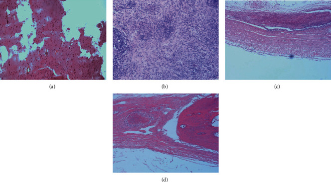
The microscopic histological images of cranial tissue sections (blank or with CGF) of the two groups stained with HE, 6 and 12 weeks after operation: (a) blank 6 weeks; (b) CGF 6 weeks; (c) blank 12 weeks; (d) CGF 12 weeks.

The microscopic histological images of cranial tissue sections (blank or with CGF) of the two groups stained with HE, 6 and 12 weeks after operation: (a) blank 6 weeks; (b) CGF 6 weeks; (c) blank 12 weeks; (d) CGF 12 weeks.