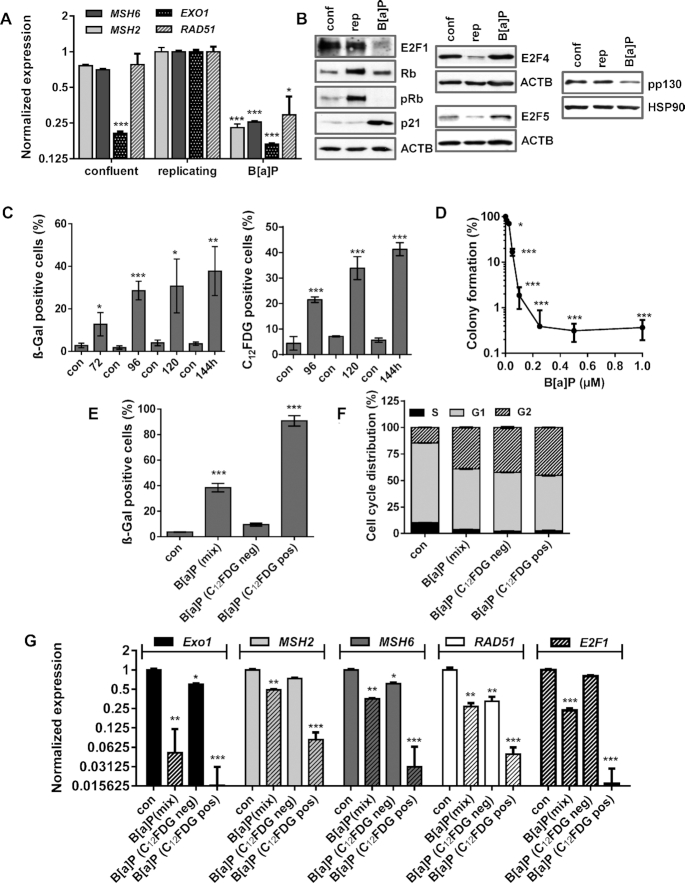
Figure 6.
(A/B) MCF7 cells were kept in confluency for one week and replicating cells were either non-exposed or exposed to 1 μM B(a)P for 72 h. (A) Expression of MSH2, MSH6, EXO1 and RAD51 was detected by qPCR. (B) Expression of E2F1, Rb, p21, E2F4, E2F5 and pp130 was detected by immunoblotting. β-Actin and HSP90 were used as internal loading control. (C) Time-dependent induction of senescence upon B[a]P was measured by microscopical detection of ß-Gal positive cells (left panel) and FACS-based detection of C12FDG positive cells (right panel). (D) Clonogenic survival upon B[a]P was measured by the colony formation assay. (E–G) MCF7 cells were exposed to 1 μM B(a)P for 120 h. C12FDG positive and negative cells were separated by FACS. As comparison, non-exposed and B[a]P-exposed cells were included. (E) Frequency of senescence was determined by β-Gal staining in non-exposed cells (con), non-sorted B[a]P-exposed cells (mix), sorted C12FDG positive and negative cells. (F) Cell cycle distribution was measured using PI staining and flow cytometry. (G) Expression of EXO1, MSH2, MSH6 and RAD51 was measured by qPCR in non-exposed cells (con), non-sorted B[a]P-exposed cells (mix), sorted C12FDG positive and negative cells. (A, C–G) Experiments were performed in triplicates and differences between treatment and control were statistically analyzed using Student's t test (non labeled = non-significant, *P< 0.1 **P< 0.01, ***P< 0.001). (F) Concerning cell cycle distribution, in non-sorted B[a]P-exposed cells (mix), sorted C12FDG positive and negative cells a significant (**) decrease of cells in G1 and increase in the G2-phase was observed.

