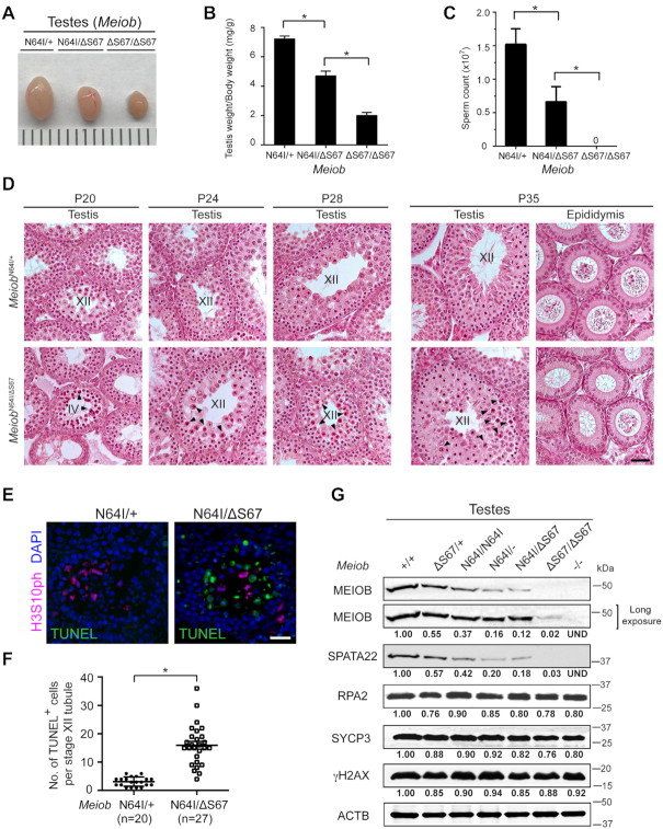
Figure 4.
Meiob N64I/ΔS67 males exhibit age-dependent meiotic defects in juvenile spermatogenesis. (A) Testis size of adult (8-week-old) males. (B, C) Testis weight (B) and sperm count (C) of adult (8-week-old) males. n, number of males. Data are shown as mean ± S.D., *P < 0.05 by Student's t-test. (D) Histological analysis of testis and epididymis at P20, P24, P28 and P35. Stage IV and stage XII tubules are labeled. Apoptotic spermatocytes are marked by arrowheads. Scale bar, 50 μm. (E, F) TUNEL analysis of MeiobN64I/+ and MeiobN64I/ΔS67 testes at P24 (E) and quantification of TUNEL-positive spermatocytes (F). n, the number of TUNEL-positive stage XII tubules (TUNEL-negative stage XII tubules were excluded). Three mice per genotype were examined. *P < 0.05 by Student's t-test. Scale bar, 20 μm. (G) Western blot analysis of MEIOB, SPATA22, and RPA2 in P20 testes. The levels of SYCP3 and γH2AX are constant across samples. Relative band intensity (normalized to ACTB) is shown.

