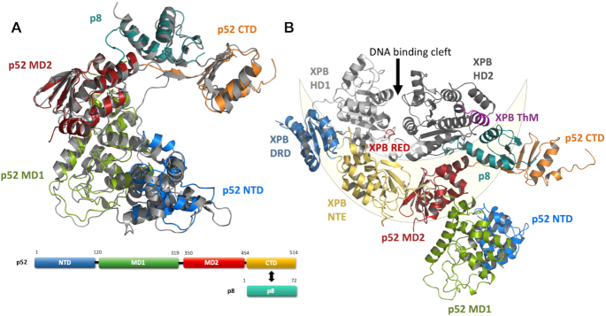
Figure 1.
Structure and domain architecture of ctp52 and ctp8. (A) Structure and domain architecture of full length ctp52 and ctp8. Ctp52 is comprised of four domains: NTD (blue), MD1 (green), MD2 (red) and CTD (orange). Ctp8 is shown in mint green. Ctp8 and ctp52 CTD adopt the same fold. The domains have been rearranged to fit to the p52 conformation found in the cryo-EM structure of human TFIIH (shown in grey). (B) Ctp52 and ctp8 modelled into the TFIIH cryo-EM structure from Greber et al. (26) from which only XPB, p52 and p8 are shown. The lunate-like ring that encircles the XPB helicase domains is schematically depicted in light yellow.

