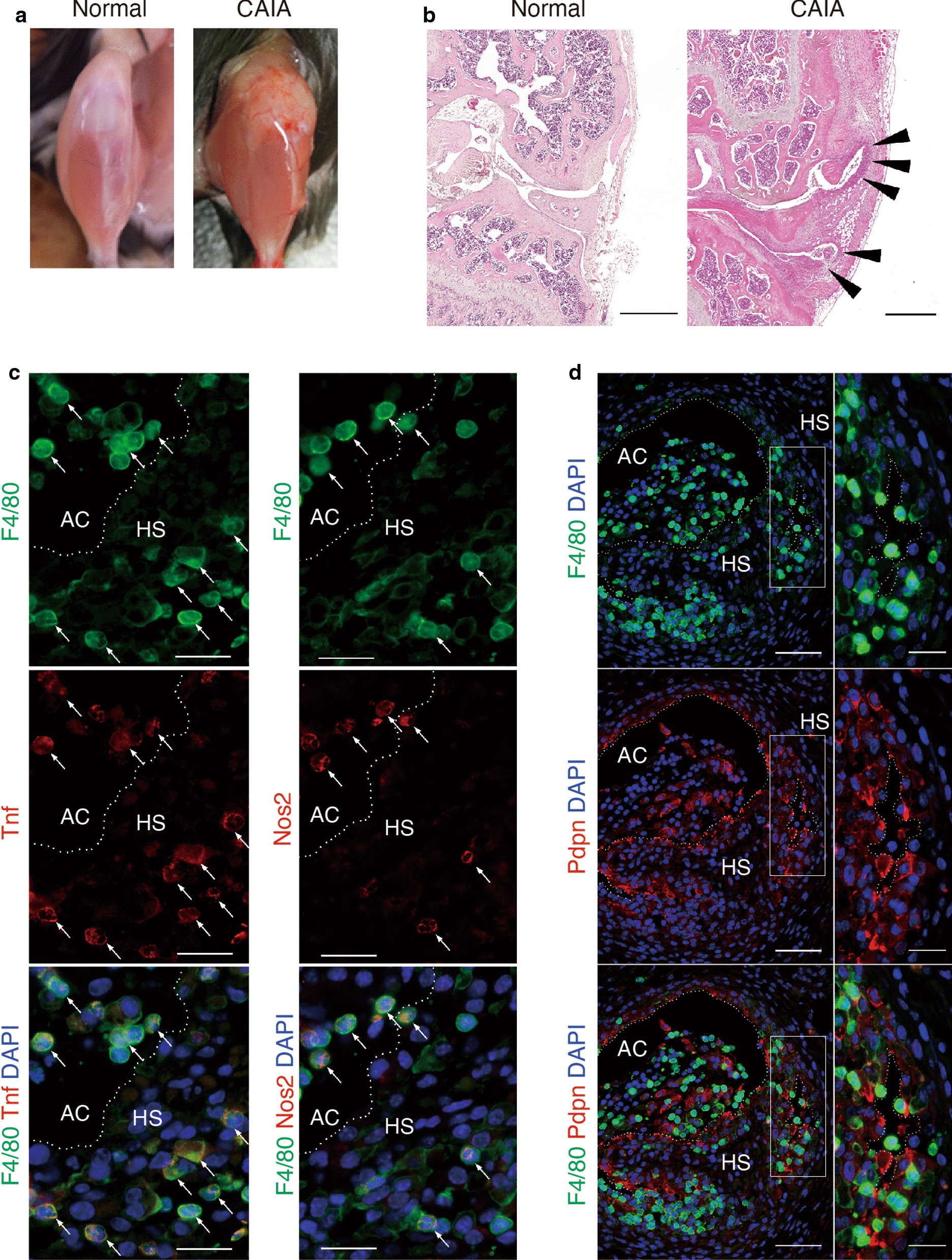
Fig. 1.

Histological analysis of knee joint tissue from CAIA mice. a Appearance of wild type mice with or without experimental arthritis (CAIA). b Sections of normal (left) and CAIA (right) knee joints stained with Hematoxylin and Eosin showing hyperplastic synovium (HS) in the joint (arrow head). Scale bar represents 500 μm. In immunofluorescent staining c F4/80-positive (green) cells were observed in the HS and articular cavity (AC). The inflammatory macrophage markers Nos2 and Tnf (red) co-localized in F4/80high-positive cells (arrow, yellow). Scale bar represents 20 μm. d Synovial cells were localized randomly in the HS. Panels on the right are high magnification images of boxed areas in the left panels. Podoplanin (Pdpn)-positive (red) cells were also observed in the HS. Scale bar represents 50 μm (left panel) and 20 μm (right panel). Histological data were technically replicated more than 2 times
