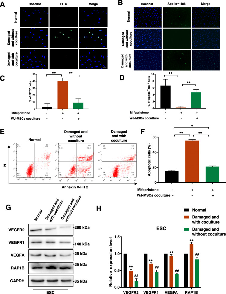
Fig. 1.
Damaged ESCs were repaired by WJ-MSCs in vitro. a TUNEL assay to detect apoptosis of ESCs in three groups. Scale bar, 20 μm. b EdU assay to detect proliferation of ESCs in three groups. Scale bar, 40 μm. c, d Quantitative analysis of TUNEL assay and EdU assay. Data in panel are shown as means ± standard deviation (n = 3). **P < 0.001. e, f ESCs in three groups were stained with Annexin V-FITC and PI, and the percentage of apoptotic cells was detected by flow cytometry. Data in panel are shown as means ± standard deviation (n = 3). *P < 0.05, **P < 0.001. g, h VEGFA, VEGFR1, VEGFR2, and RAP1B protein expression of ESCs in three groups by western blot assay. Data in panel are shown as means ± standard deviation (n = 3). **P < 0.01 compared with the damaged and without coculture group, ##P < 0.01 compared with the normal group

