Figure 2.
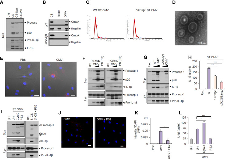
Outer membrane vesicles from Salmonella typhimurium activate inflammasome signaling via receptor-mediated endocytosis. (A) Immunoblots from the culture supernatants of mouse BMDMs treated with S. typhimurium CS, or supernatant (CS-Sup) or pellet (CS-Pel) from ultracentrifugation (150,000 × g, 18 h) of S. typhimurium CS. (B) Immunoblots from wild-type (WT) or ΔfliC–fljB S. typhimurium CS, filtrates from Centricon concentration ( Supplementary Figure S3A ) and the extract of OMVs. (C) Nanoparticle Tracking Analysis of OMVs isolated from WT or ΔfliC–fljB S. typhimurium. (D) Transmission electron microscopy of OMVs isolated from wild-type S. typhimurium. Scale bar, 100 nm. (E) Representative confocal microscopy images of BMDMs treated with Vybrant Dil-labeled PBS or S. typhimurium OMVs. Scale bar, 20 µm. (F) Immunoblots from mouse BMDMs treated with OMVs isolated from S. typhimurium SL1344 or 14028s (0.5 or 5 µg/ml) for 8 h or treated with Pam3CSK4 (1 µg/ml, 4 h), followed by the transfection of LPS (2 µg/ml, 5 h) using a DOTAP (DT). (G) Immunoblots from mouse BMDMs treated with WT or ΔfliC–fljB S. typhimurium (SL1344) OMV (1 or 5 µg/ml, 8 h). (H) Quantification of IL-1β in the culture supernatants of mouse BMDMs treated with WT, ΔfliC–fljB, or ΔfliC–fljB–prgJ S. typhimurium (SL1344) OMVs (5 µg/ml, 8 h) (n = 7, one-way ANOVA). (I) Immunoblots from mouse BMDMs treated with S. typhimurium OMV (5 µg/ml, 6 h) in the presence of cytochalasin D (5 µM) or Pitstop 2 (PS2, 10 µM), or treated with S. typhimurium CS (1/20) in the presence of PS2 (10 µM) pretreatment (10 min before Salmonella CS treatment). (J) Representative confocal microscopy images of BMDMs treated with Vybrant Dil-labeled S. typhimurium OMVs (5 µg/ml, 6 h) in the presence of Pitstop 2 treatment (10 µM, 30 min pretreat). Scale bar, 20 µm. (K) Relative Dil fluorescence intensity per DAPI signals of BMDMs as treated in (J). (n = 6, one-way ANOVA). (L) Quantification of IL-1β in the culture supernatants of mouse BMDMs treated as in (I, left panel). (n = 3, one-way ANOVA). Culture supernatants (Sup) or cellular lysates (Lys) were immunoblotted with the indicated antibodies. Data were expressed as the mean ± SEM. Asterisks indicate significant differences compared with S. typhimurium CS-treated group. (*P < 0.05, ***P < 0.001).

