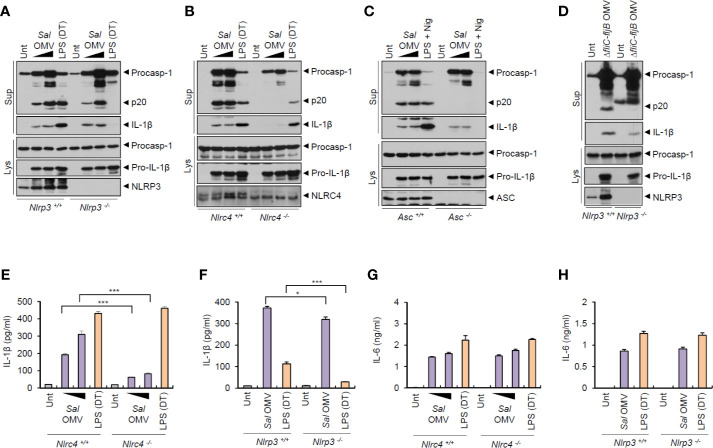
Figure 3.
Salmonella-released outer membrane vesicles promotes NLRC4-dependent inflammasome activation. (A, B) Immunoblots from Nlrp3 +/+ or Nlrp3 −/− (A) or Nlrc4 +/+ or Nlrc4 −/− (B) mice BMDMs treated with S. typhimurium OMVs (3 or 10 µg/ml, 6 h, A; 5 or 12.5 µg/ml, 6 h, B), or treated with Pam3CSK4 (1 µg/ml, 4 h), followed by the transfection of LPS (1 µg/ml, 6 h) using a DOTAP (DT). (C) Immunoblots from Asc +/+ or Asc −/− immortalized BMDMs treated with S. typhimurium OMVs (5 or 12.5 µg/ml, 6 h), or primed with LPS (1 µg/ml, 6 h), followed by the treatment with nigericin (5 µM, 40 min). (D) Immunoblots from Nlrp3 +/+ or Nlrp3 −/− mice BMDMs treated with ΔfliC–fljB S. typhimurium OMVs (10 µg/ml) for 6 h. (E, F) Quantification of IL-1β in the culture supernatants of Nlrc4 +/+ or Nlrc4 −/− (E) or Nlrp3 +/+ or Nlrp3 −/− (F) mice BMDMs treated with S. typhimurium OMVs (1 or 7 µg/ml, E; 10 µg/ml, F) for 8 h, or treated with Pam3CSK4 (1 µg/ml, 3 h), followed by the transfection of LPS (2 µg/ml, E; 1 µg/ml, F) for 6 h. (n = 3) (G, H) Quantification of IL-1β in the culture supernatants of Nlrc4 +/+ or Nlrc4 −/− (G) or Nlrp3 +/+ or Nlrp3 −/− (H) mice BMDMs treated with as same as in (E, F). (n = 3) Culture supernatants (Sup) or cellular lysates (Lys) were immunoblotted with the indicated antibodies. Data were expressed as the mean ± SEM. Asterisks indicate significant differences compared with the group in the Nlrp3 +/+ cells. (*P < 0.05, ***P < 0.001).

