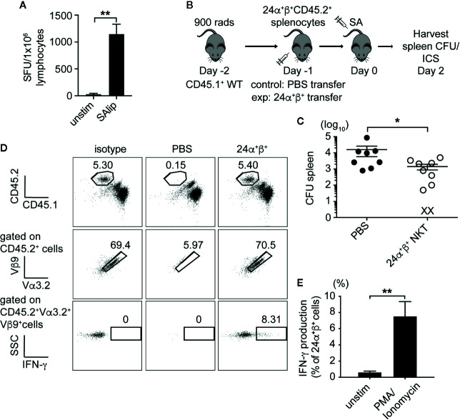
Figure 7.
Type II NKT cells protect mice from systemic SA infection. (A) IFN-γ ELISPOT of naïve 24α+β+ lymphocytes from liver co-cultured with B6 DCs unpulsed or pulsed with total SA lipids (SAlip) (10 µg/ml). (B) Schematic of type II NKT cell adoptive transfer experiment. (C) Bacterial CFU in the spleen of recipient mice at 2 dpi, PBS= control group (D, E). Representative FACS plots of adoptively transferred 24α+β+ NKT cells from spleen of 2 dpi recipient, % IFN-γ production after stimulation with PMA/Ionomycin or unstimulated (2 h + 4 h BFA), quantified in E (N=8–10 mice/group). Statistical analysis: (A) one-way ANOVA, (C) Mann-Whitney test, (E) 2-way ANOVA. *p < 0.05; **p < 0.01.

