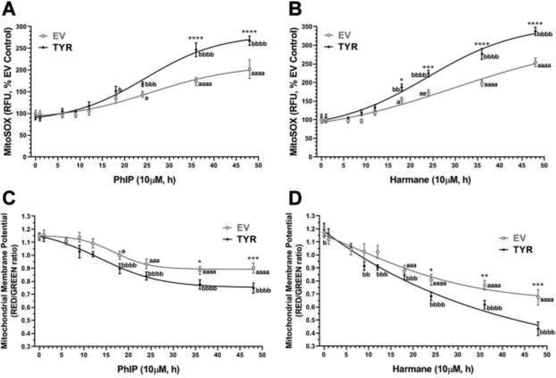
Figure 1: HAAs increase mitochondrial superoxide and reduce mitochondrial membrane potential production in a time-dependent manner in neuromelanin forming SH-SY5Y cells.

Galactose-supplemented SH-SY5Y neuroblastoma cells were transfected with EV or TYR. After 48h of transfection, cells were treated with either 10 μM HAR or 10 μM PhIP in a time-dependent manner from 1 h to 48 h. A, B. Mitochondrial superoxide (MitoSOX) levels analyzed for each time-point post-treatment with PhIP or HAR, respectively. C, D. Time-dependent changes in mitochondrial membrane potential (Δψm) post-treatment with PhIP or HAR, respectively. The data are represented as mean ± SEM for n = 6, analyzed by two-way ANOVA followed by Sidak’s post-hoc test. ap<0.05, aaap<0.001 and aaaap<0.0001 compared EV non-treated control group; bp<0.05, bbp<0.01, bbbp<0.001 and bbbbp<0.0001 compared TYR non-treated control group; while, *p<0.05, **p<0.01, ***p<0.001 and ****p<0.0001 demonstrate significant difference between EV versus TYR-transfected SH-SY5Y cells at given concentration.
