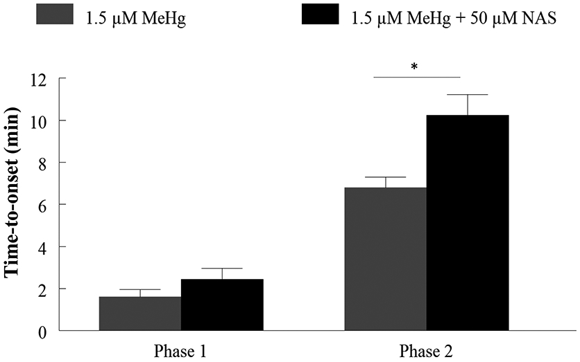
Figure 11. Acute MeHg exposure-induced increases of Ca2+i in hiPSC-MNs are mediated in part by Ca2+-permeable AMPARs.

MeHg-induced increase in fluorescence time-to-onset is delayed by exposure to 50 μM NAS, a Ca2+ permeable AMPAR antagonist, confirming that this receptor is contributing to MeHg-induced increases in [Ca2+]i in hiPSC-MNs. The asterisk (*) indicates a significant difference between 1.5 μM MeHg alone vs 1.5 μM MeHg + 50 μM NAS (p ≤ 0.05) as determined by Two-Way ANOVA and Bonferroni’s post hoc test. Values are represented as mean ± SEM (n = 3).
