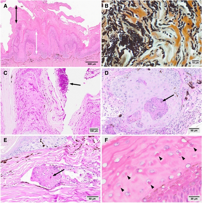
Figure 2.
Histopathological features of cutaneous lesions from free-living Australian lizards with Nannizziopsis barbatae-associated dermatomycosis (A) severe hyperkeratosis (black double pointed arrow), papillary epidermal hyperplasia (white double pointed arrow) (case EWD009; H&E). (B) Abundant fungal hyphae and arthroconidia, morphologically consistent with Nannizziopsis spp. (Case EWD009; GMS) (C) surface tuft of arthroconidia (arrow) (case EWD009; PAS). (D) Dysplastic invaginating epithelium with intralesional fungal hyphae (arrow) (case TRH001; PAS). (E) Perivascular dermal granuloma with intralesional hyphae (arrow) in the sole animal with deep dermal mycosis (case EWD008; PAS). (F) Epidermal intracytoplasmic inclusion bodies (arrowheads) (Case EWD009; H&E).

