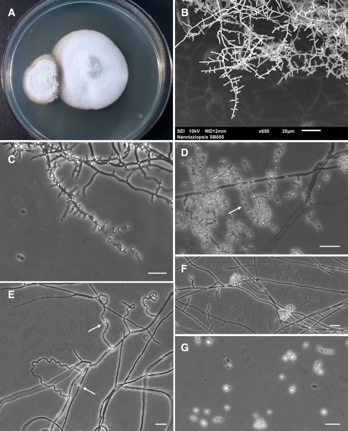
Figure 3.
Morphological characteristics of Nannizziopsis barbatae, isolated from cutaneous lesions of free-living Eastern water dragons and incubated on PDA at 30 °C. (A) Colony morphology on PDA at 21 days. (B) Scanning electron micrograph showing branched fungal hyphae bearing aleurioconidia. (C) Microscopic morphology showing pyriform to clavate, mainly sessile aleurioconidia, 2.5–10 μm in length and 1.5–3 μm in width, born along septate branching hyphae and occasionally subtended by swollen cells. (D) Fission arthroconidia, 2.8–8.3 μm in length and 1.8–3.3 μm in width. (E) Abundant undulate hyphae, characteristic of Nannizziopsis species8, and intercalary chlamydospores (arrows). (F) Ascomatal initials and (G) budding cells were occasionally present. Bars 20 μm.

