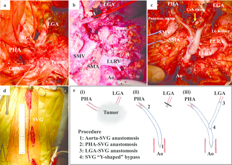
Fig. 3.
Intraoperative findings. a The tumor involved the surrounding arteries. b Plan to reconstruct arteries after dissecting the pancreas. c Image obtained after tumor resection and reconstruction of PHA and LGA. d Harvesting the SVG. e The details of the procedures in grafting. e-(i), (ii): after the aorta and the SVG graft was anastomosed, the PHA and the SVG graft was anastomosed. e-(iii): another SVG graft was anastomosed to the LGA. Finally, the first and second SVG were anastomosed together to form a Y-shape

