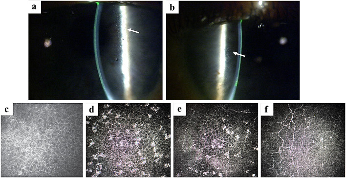
Fig. 4.
Case report representative slit lamp and in vivo confocal microscopic images of MECs. MECs microcyst-like epithelial changes. Slit lamp microscopic images of the right (a) and left (b) eyes, demonstrating MECs (arrows). These lesions are best visualized at medium to high magnification. Retroillumination or indirect illumination can be helpful. In vivo confocal microscopic image from the same patient (c–f) demonstrating hyperreflective opacities within the corneal epithelium. These opacities were noted to be most prominent in the wing cells (d) and basal cells (e), as compared with the superficial cells (f). They were not visualized within the anterior stroma (f) or endothelium (not shown)

