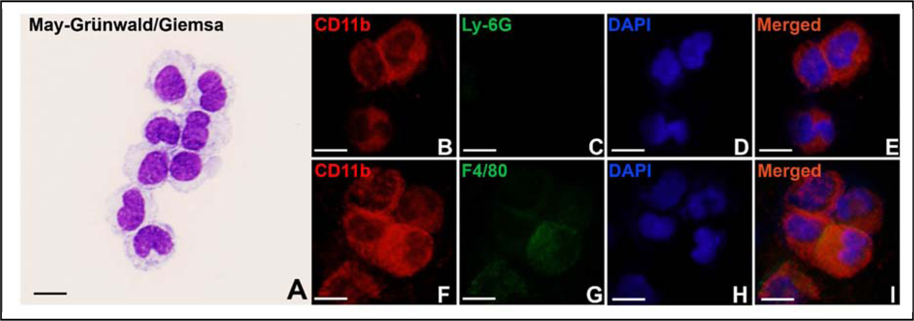
Fig. 1. Isolated monocytes.

Monocytes were isolated from mouse peripheral blood and stained with May-Grünwald and Giemsa stains (A), or with anti-CD11b (B&F) and anti-Ly-6G (C) or anti-F4/80 (G) antibodies. DAPI stains the whole nucleus of a cell (D&H). E&I are merged images. Scale bar represents 10 μm for all panels.
