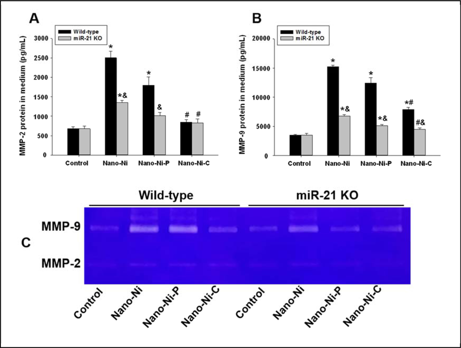
Fig. 6. MMP-2 and MMP-9 protein levels and activities in the monocyte culture medium.

Monocytes were isolated from peripheral blood of wild-type and miR-21 KO mice and treated with 30 μg/mL of Nano-Ni or with either Nano-Ni-P or Nano-Ni-C with same molar concentration of Ni as Nano-Ni for 24 h. Cells treated with physiological saline were used as the control. MMP-2 or MMP-9 protein level was determined by Mouse MMP-2 or MMP-9 PicoKine™ ELISA Kit (A-B), while MMP-2 and MMP-9 activities were determined by gelatin zymography assay (C). Data are shown as mean ± SE (n=3). * p<0.05 vs. Control; # p<0.05 vs. Nano-Ni group; & p<0.05 vs. wild-type group with the same treatment.
