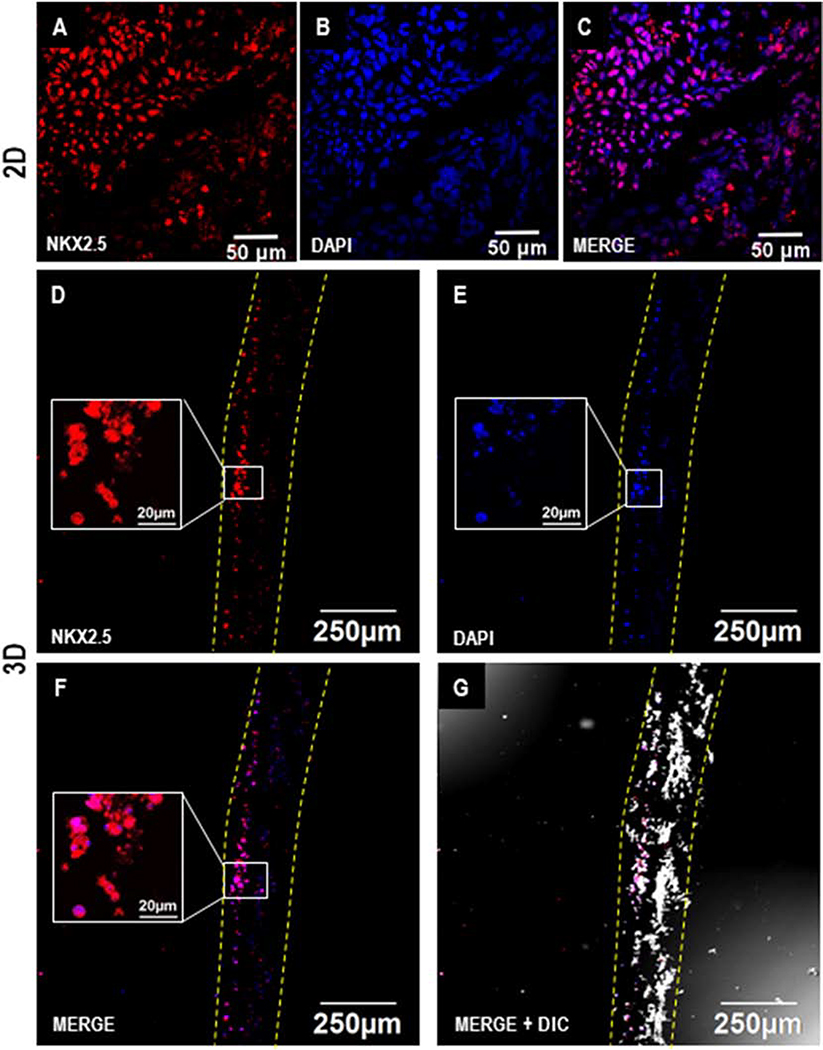
Figure 5.
Immunofluorescence analysis of cardiac differentiation of hiPSCs in 2D and 3D cultures on D7. Confocal images showing the expression of cardiac progenitor marker NKX-2.5 on D7 during cardiac differentiation of hPSCs in 2D (A-C) and 3D (D-G). Scale: A-C: 50 μm; D-G: 250 μm; inset figures (D-F): 20 μm. Dotted yellow lines represent the coaxial scaffold.

