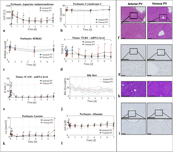
Figure 3.
Injury markers and liver function in experimental groups. Arterial PV group blue circles, n = 5 experiments, venous PV group red triangles, n = 5 experiments. (a–c) Injury marker release in perfusate shown for AST (a), cytochrome c (b) and 8-OHdG (c). (d,e) Representative staining showing integrity on H&E staining (g) and preserved glycogen stores in PAS staining (h) after one week of perfusion. (f) Representative immunohistochemistry staining for Caspase 3 showing absence of relevant apoptosis on perfusion day 7. (d) Macrophages were not activated in both groups as shown with TLR4 at mRNA level. (e) Endothelial cell activation at mRNA level expressed with ICAM-1. (i) similarly to ICAM-1, von Willebrand Factor immunohistochemistry staining showed absence of relevant endothelial cell activation. (j) Bile flow was constantly present in both experimental groups for one week. (k,l) Livers cleared lactate (k) and maintained albumin level (l) in perfusate. P value * < 0.05, ** < 0.01, *** < 0.001. ns not significant.

